Welcome to VGMorph GmbH
Definite 4D-Morphometry

VGMorph qualified for ‘Successful R & I in Europe’
We are delighted to have qualified as VGMorph for the ‘Successful R & I in Europe’ meeting in Düsseldorf.

Participation 2. Startup Night in Frankfurt on March 27, 2025
We are very happy to have been part of the 2nd Startup Night in Frankfurt am Main on March 27, 2025
What is Voxel-Guided Morphometry (VGM)?
4D
precise
efficient
cutting edge
Voxel-Guided Morphometry (VGM) is a method for accurate morphometric analysis of brain changes based on MRI scans.
By comparing different individuals (‘cross-sectional’ analysis) or serial examinations of the same individual (individual longitudinal analysis), individual examinations and also group analyses can be performed.
The results are visualized, absolutely quantified and statistically evaluated.
Our mission
New dimensions in brain morphometry
We are rethinking the analysis of brain imaging data.
Voxel-Guided Morphometry (VGM) offers an innovative approach to analyze volumetric data of the brain and thus to accurately visualize structural changes of the brain. This provides a significantly improved understanding of change processes in the brain.
By using VGM, a forward-looking diagnostic approach can be achieved with the aim of sustainably improving the health and quality of life of patients.
Who benefits from VGM?
Users
Pharmaceutical companies
To assist in clinical trials in all clinical phases in assessing the efficacy of new drugs or therapeutic procedures
Universities and academic research institutions
Cooperations in projects to study structural changes in the brain - in humans and/or animals
Clinics/Practices
Assist in the evaluation of serial patient MRIs of patients with neurological or neurosurgical conditions.
MRI/CT manufacturing companies and app developers
Options for integrating the VGM software into diagnostic devices
Strengths of VGM:
Powerful & informative
- High spatial resolution of analyses (approx. 0.8 mm when using a 3T scanner)
- Automated examination of the entire brain volume
- High reproducibility through automation
- Absolute quantification
- Possibility of intraindividual and group analyses
- Statistical, spatially resolved evaluability
- Fast examination, also suitable for large studies
Benefits for users
New insights through better data
Quality of results
- Better results through individual morphometry
- Three-dimensional representation of changes throughout the entire brain volume
Gain in knowledge
- New insights through improved precision. By means of VGM, results are obtained that in part elude visual analysis. This supports radiologists / neurologists / neurosurgeons in their diagnostic work
Profitability
- More accurate results enable more targeted use of financial resources
Speed and quality of service
- After receiving the MRI scans, we take care of everything else. You receive fully visualized, quantified and statistically evaluated results
Diagnostics
- Support for clinically active radiologists, neurologists and neurosurgeons in optimized diagnostics based on MR tomographic examinations
Pharmacological studies
- Structural effects of pharmacological therapies can be detected at the individual level
- VGM detects possible influences of pharmacological therapies on the brain structure of individual subjects or groups
Science
- VGM supports neuroscience research in detecting morphological changes of the brain under various conditions
- Neurological diseases such as multiple sclerosis, Parkinson's disease, dementia, tumors, stroke, etc
- Physiological changes due to adolescence, aging, learning etc
Our cooperation partners
With the best recommendations
Contributions to scientific publications (selection)
MRI-based morphometric structural changes correlate with histopathology in experimental autoimmune encephalomyelitis
Magnetic resonance imaging (MRI) and neurohistopathology are important correlates for evaluation of disease progression in multiple sclerosis (MS).
Venous Diameter Changes in Chronic Active Multiple Sclerosis Lesions
To investigate the temporal evolution of venous diameter in chronic active and nonenhancing shrinking multiple sclerosis (MS) lesions in a longitudinal magnetic resonance imaging (MRI) study including susceptibility‐weighted images (SWI).
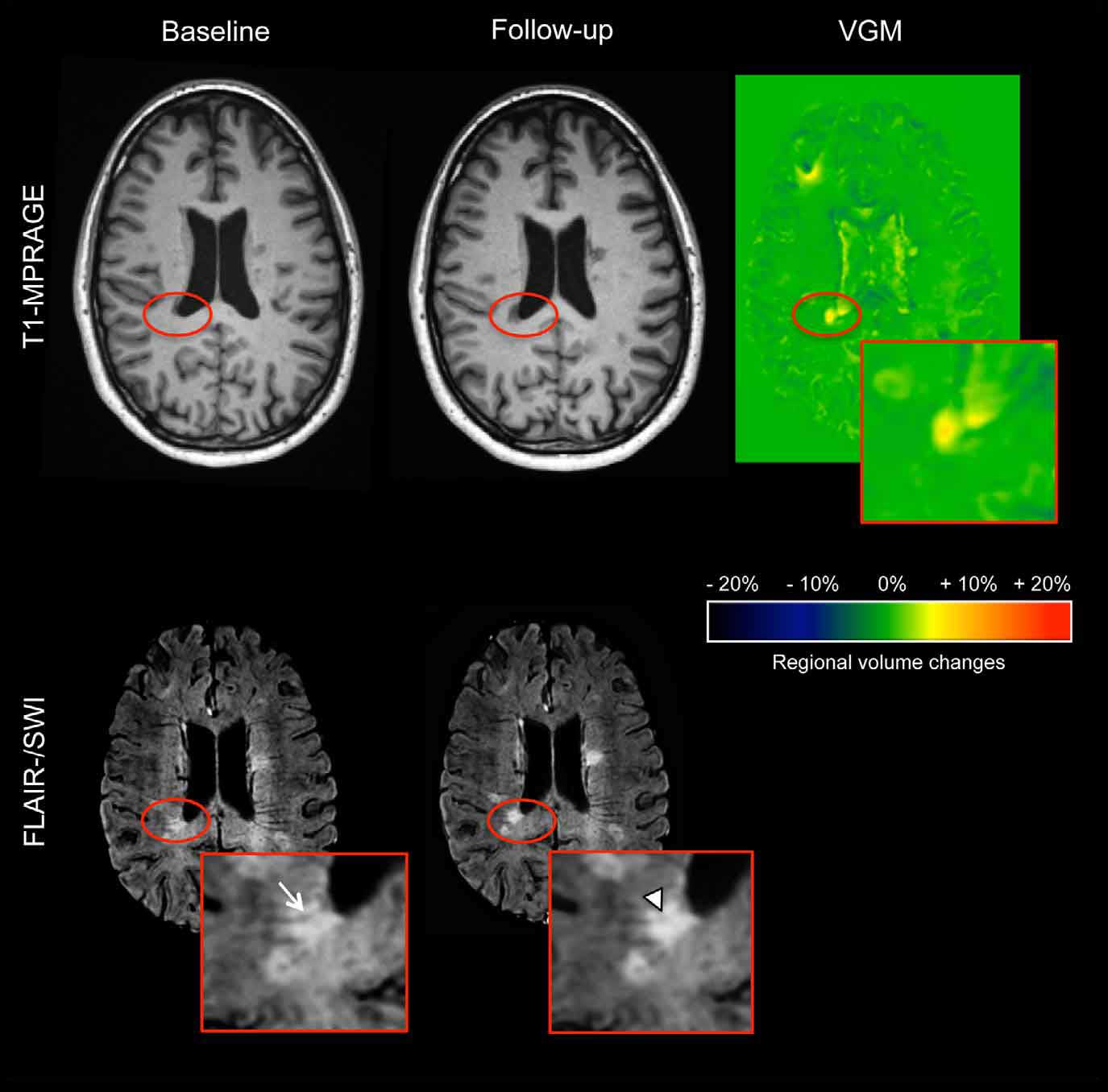
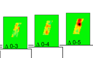
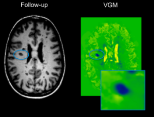
Individual Assessment of Brain Tissue Changes in MS and the Effect of Focal Lesions on Short-Term Focal Atrophy Development in MS: A Voxel-Guided Morphometry Study
We performed voxel-guided morphometry (VGM) investigating the mechanisms of brain atrophy in multiple sclerosis (MS) related to focal lesions.